 It’s really squeezed my energy to think and at last to get a diagnose of a very rare tumor of central nervous system located at the frontal lobe.
Neurosurgeon said that the tumor was en-plaque, maximum spread to the duramater with no penetration to the hemisphere. On X-ray examination there were no bone destructions, only bone enhanched. And he assumed it to be a benign tumor, eg. osteoma.
 Under a light microscope, I found some interesting clues lead to malignancy. At last I asked a second opinion to the CNS-pathologists at hospital Cipto Mangunkusumo (Central National Hospital in Jakarta).
Let’s see what they say…
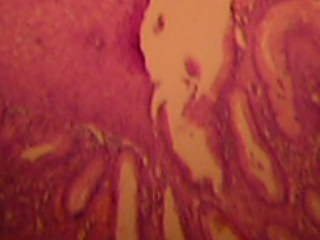
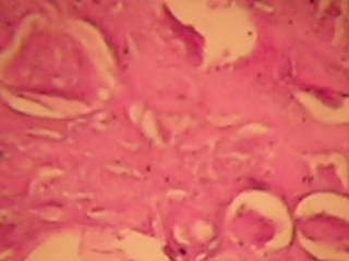
One week later, I received the answer from Cipto Mangunkusumo Hospital in Jakarta. They made Differential Diagnoses as : 1. Intramedullary Hemangiopericytoma and 2. Meningioma mixed type : angiomatous and clear cell-secretoric. Is it ? I am not 100 % satisfied with the answer. The microscopic features as we can see in the two pictures attaching to this article. Hope the patient is doing well until now..
Popularity: unranked
 Dr. Sukma Merati is founder and owner of Riau Pathology Center in Pekanbaru, Riau. Dr. Merati has had various international experience and training, including as a fellow doctor at The Mount Sinai Hospital in New York City, NY, USA (2000-2002).
February 23rd, 2008 at 5:34 pm
Dr. Devi, it’s great to see your comment on my blog. Thanks. You really are a dynamic and always eager to see more progresses in our field of medicine. I will write more about everything in my mind, until nothing left in there…
February 21st, 2008 at 10:58 am
dr.Sukma, SpPA..congratulation for your new website !! I hope this website..can give information not only about anatomical pathology..ok can’t wait to read your next blog?? or next article??