Lately, I think I’ve short of time. So many cases that I have to handle, to be examined first by me and later on be processed by my technician which at last I have to look at the microscope to see and finally reveal the diagnoses.
 The first case was a radical mastectomy from a 37 years old woman with breast cancer. One week ago this patient was done a biopsy on a 6 centimeters lump of her right breast, which already diagnosed with Invasive Ductal Carcinoma. The specimen was measured 15x 15x 3 cm, with nipple and skin attached on it. I have to see diligently, if all the margins of the operation : cranial, caudal, medial, lateral and base of the section were all free from tumor cells. So did to the possibility of vascular invasion or tumor emboli and lymph nodes dissected from her axilla or her right armpit. I found there were massive vascular invasions and infiltration of tumor cells to all the twelve lymphnodes of her right armpit.
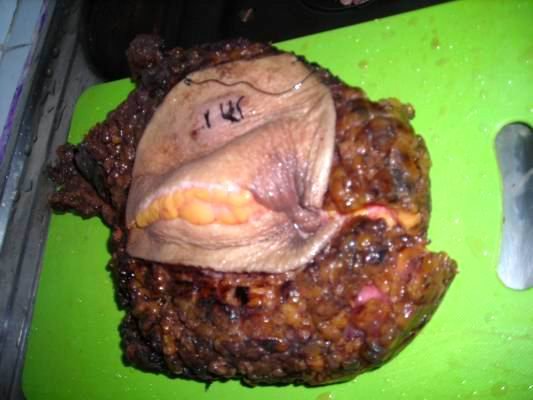
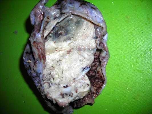 The second case was a huge ovary cancer of a 43 years old woman. The specimen measured 16x 9x 7 cm, with a hard portion almost to the entire specimen. The outer surface was glistening, greyish-white. On slicing, I found an enormous greywhitish fleshy mass with rubber to hard consistency which later on I’ve made a diagnosis of Solid Embryonal Carcinoma of the Ovary. Under light  microscope the morphology was almost like seminoma, but without lymphocytes infiltration in the stroma. I think the malignancy of this Solid Embryonal Carcinoma of the ovary is moderate. This patient has a bleak prognosis, because there’s already metastases to the peritoneum with adhesions to her bowels and many nodules on her liver..
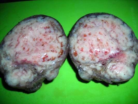 The third case was a huge fibroid tumor of the womb or Benign Uterine Leiomyoma . The specimen measured 16 centimeters in diameter. On cut surfaces it looked fleshy fresh with bleeding spots. This specimen from a woman, 42 years of age. Imagine how big was her stomach before the operation taken to throw out her solid benign tumor of her womb which she has had for 3 years’ time.
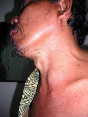 The fourth case was a male patient, 56 years of age with a lump on his thyroid gland left lobus measuring 4 centimeters in diameter. This lump was known only 3 months before visiting my laboratory. Rubber consistency and a bit painful on palpation. He admitted that he got fever and difficulty in swallowing and feels short of breath. I’ve done a fine needle aspiration biopsy to his lump. And ultimately my diagnosis was Subacute Thyroiditis or de Quevain’s Thyroiditis. Lucky for him that his tumor need not to be operated, and will be fine and back to normal within 2-3 weeks time with only antibiotic and steroid tappering off therapy.
Popularity: unranked
 Dr. Sukma Merati is founder and owner of Riau Pathology Center in Pekanbaru, Riau. Dr. Merati has had various international experience and training, including as a fellow doctor at The Mount Sinai Hospital in New York City, NY, USA (2000-2002).
August 15th, 2011 at 1:36 pm
halo Dr.Sukma,
kenalkan saya devi,
saya ingin bertanya tentang masalah kanker, saya mahasiswa yang sedang mengerjakan tugas akhir, dan sedang mencari tahu tentang botol minum kemasan plastik PET.
saya menemukan beberapa artikel di Internet bahwa penggunaan botol plastik kode 1 / 2 secara terus menerus dan terkena panas / sinar matahari, dapat menyebarkan zat karsinogen yang dapat menyebabkan kanker, apakah hal ini benar?
terima kasih sebelumnya dok
March 23rd, 2010 at 11:57 pm
hai Muthy,
Kunjungi dokter umum agar dirujuk ke dokter spesialis PA (patologi anatomik) untuk dilakukan FNAB atau biopsi jarum halus terhadap benjolan itu. Hasil FNAB selanjutnya dibawa ke dokter spesialis penyakit dalam agar diberikan terapi yang sesuai diagnosis biopsi tsb.
March 22nd, 2010 at 3:37 am
Dok, d bagian telinga bwh saya dpn n belakan mngalami pembengkakan, sdh 3 hr brtambah bsr. Berbahaya g dok? N gmn cr ngatasinya
September 9th, 2009 at 9:21 pm
Halo Wiwi,
Yang jelas sepertinya anda terkena migrain. Biasanya disebabkan banyak pikiran, stress dan permasalahan pelik.
Sebaiknya dibereskan semua masalah yang ada, sambil minum obat migrain yang bisa dibeli di warung/toko obat.
September 9th, 2009 at 12:48 pm
Dear Dr. Sukma,
Dok, ud 2 hari ini saya merasakan sakit kepala sebelah kiri, rasa nyeriny begitu menusuk. kira2 10 menit rasa nyeri itu berulang terus. Nyeriny sampai terasa dikulit kepala s/d kekulit dekat telinga saya Dok.
Tadi pagi rasa nyeri itu juga terasa di bagian kanan jg.
Yang mau saya tanyakan apakan itu hanya sakit kepala biasa dok, atau gejala2 penyakit lainny?
Terima kasih,
Wiwi
April 28th, 2009 at 6:39 pm
Salam kenal dok, sebelumnya saya ingin mengataka bhw blog dokter sagat bagus dan penuh pengetahuan
Dok sy mau tanya… kasus patologi anatomi apa ya yang sekarang hangat dan kira-kira mampu saya teliti untuk KTI saya